


Free ARDMS (SPI) Certification Sample Questions with Online Practice Test
SPI Certification Study Guide Pass SPI Fast
ARDMS SPI Exam Syllabus Topics:
| Topic | Details |
|---|---|
| Topic 1 |
|
| Topic 2 |
|
| Topic 3 |
|
| Topic 4 |
|
| Topic 5 |
|
NEW QUESTION # 100
Which color control was adjusted in color bar A to produce color bar B?
- A. Scale
- B. Baseline
- C. Map
- D. Invert
Answer: A
Explanation:
The color bar on a Doppler ultrasound display indicates the range of velocities that the system can detect and display. In color bar A, the scale is set to a higher maximum velocity (64 cm/s), while in color bar B, the scale is set to a lower maximum velocity (16 cm/s). Adjusting the scale (or velocity range) changes the upper and lower limits of the velocities displayed, which affects the sensitivity of the Doppler system to detect flow velocities. Lowering the scale allows for better visualization of lower velocities, but it may also increase the likelihood of aliasing for higher velocities.
American Registry for Diagnostic Medical Sonography (ARDMS). Sonography Principles and Instrumentation (SPI) Examination Review Guide.
NEW QUESTION # 101
Which artifact is seen as a result of an increase in echo amplitude in the tissue located distal to an anechoic structure?
- A. Comet tail
- B. Enhancement
- C. Reverberation
- D. Mirror image
Answer: B
NEW QUESTION # 102
Which artifact results from decreased attenuation?
- A. Comet tail
- B. Enhancement
- C. Reverberation
- D. Ringdown
Answer: B
Explanation:
Enhancement is an artifact that results from decreased attenuation. When an ultrasound wave travels through a medium with lower attenuation compared to surrounding tissues, it loses less energy. Consequently, the structures located deeper than the low-attenuation medium appear brighter on the ultrasound image. This artifact is commonly observed behind fluid-filled structures, such as cysts or the urinary bladder, where the sound waves encounter minimal resistance and thus less attenuation.References:
* ARDMS Sonography Principles and Instrumentation guidelines
* "Diagnostic Ultrasound: Principles and Instruments" by Frederick W. Kremkau
NEW QUESTION # 103
Which image demonstrates appropriate spectral Doppler gain?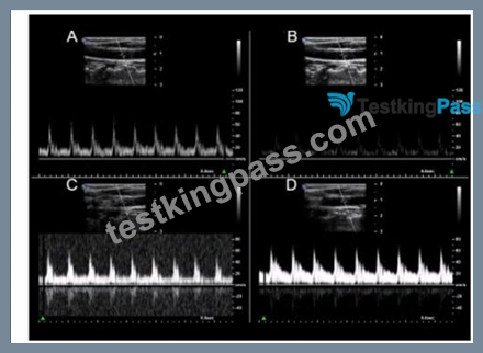
- A. Option B
- B. Option A
- C. Option C
- D. Option D
Answer: A
Explanation:
Option B demonstrates appropriate spectral Doppler gain. Appropriate gain settings ensure that the Doppler signal is adequately amplified without introducing excessive noise or artifacts. In Option B, the spectral waveform is clearly visible with distinct borders, and the background noise is minimal. In contrast, other options might show either under-gained (too little signal) or over-gained (excessive noise and signal) images, making it difficult to accurately interpret the spectral Doppler information. Reference:
ARDMS Sonography Principles and Instrumentation guidelines
"Understanding Ultrasound Physics" by Sidney K. Edelman
NEW QUESTION # 104
Which resolution can be evaluated in the area indicated by the red oval in this image of a tissue-equivalent phantom?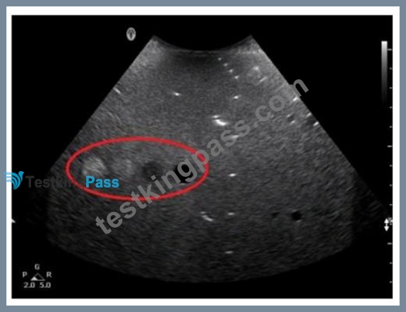
- A. Axial
- B. Contrast
- C. Elevational
- D. Lateral
Answer: A
Explanation:
The tissue-equivalent phantom image with the red oval indicates an area where axial resolution can be evaluated. Axial resolution refers to the ability to distinguish between two structures that are close together along the axis of the ultrasound beam. It is determined by the spatial pulse length (SPL) of the ultrasound wave. In phantoms, this is typically tested by observing the ability to separate closely spaced targets along the beam's path.
Reference:
ARDMS Sonography Principles & Instrumentation Guidelines
Hedrick WR, Hykes DL, Starchman DE. Ultrasound Physics and Instrumentation. 4th ed. Philadelphia, PA: Elsevier Saunders; 2005.
NEW QUESTION # 105
Which factor affects lateral resolution in ultrasound?
- A. Propagation speed
- B. Depth of penetration
- C. Wavelength
- D. Beam width
Answer: D
Explanation:
Comprehensive and Detailed Explanation From Exact Extract:
Lateral resolution is the system's ability to distinguish two structures side-by-side. It is directly determined by the beam width - the narrower the beam, the better the lateral resolution.
Principles and Instrumentation:
"Lateral resolution depends on beam width at a given depth. Narrower beams provide better lateral resolution."
* Depth of penetration influences maximum imaging depth.
* Propagation speed is largely constant in soft tissue.
* Wavelength affects axial resolution.
Therefore, the correct answer is A: Beam width.
-
NEW QUESTION # 106
Which change should be made to lower the mechanical index (MI)?
- A. Decreasing output power
- B. Lowering transducer frequency
- C. Activating tissue harmonics
- D. Increasing gain
Answer: A
Explanation:
Comprehensive and Detailed Explanation From Exact Extract:
Mechanical Index (MI) is proportional to the peak negative pressure and inversely proportional to the square root of frequency. Lowering output power directly decreases the peak pressure, thus reducing MI.
Principles and Instrumentation state:
"Mechanical index decreases with lower output power, reducing the risk of mechanical bioeffects such as cavitation." Lowering frequency (A) increases MI.
Tissue harmonics (B) improves image quality but does not reduce MI directly.
Increasing gain (D) affects displayed brightness, not acoustic power.
Therefore, the correct answer is C: Decreasing output power.
-
NEW QUESTION # 107
Which type of resolution does damping improve?
- A. Axial
- B. Contrast
- C. Temporal
- D. Lateral
Answer: A
Explanation:
Comprehensive and Detailed Explanation From Exact Extract:
Damping reduces the number of cycles per pulse, shortening pulse duration and spatial pulse length, which directly improves axial resolution.
Principles and Instrumentation:
"Axial resolution improves with shorter spatial pulse length, achieved by damping, which limits ringing of the transducer."
* Lateral resolution (A) depends on beam width.
* Contrast resolution (B) relates to dynamic range.
* Temporal resolution (C) is tied to frame rate.
Therefore, the correct answer is D: Axial.
NEW QUESTION # 108
Which statement characterizes the primary difference between image A and image B?
- A. Image A demonstrates a lower overall gain setting.
- B. Image A demonstrates a shallower field of view.
- C. Image A demonstrates a wider scale of contrast.
- D. Image A demonstrates a better axial resolution.
Answer: A
Explanation:
Comprehensive and Detailed Explanation From Exact Extract:
In image A, the structures appear darker with less overall brightness compared to image B. This indicates that the overall gain (receiver amplification) is set lower in image A, resulting in a dimmer image. Gain controls how much the returning echoes are amplified after detection.
According to Principles and Instrumentation:
"Overall gain amplifies all returning echoes equally. A lower gain setting results in a darker image, while higher gain brightens the display." Axial resolution (A) is primarily dependent on frequency and pulse length, not visible here.
Field of view (C) appears similar between both images.
Contrast scale (D) refers to dynamic range, not directly indicated here.
Therefore, the correct answer is B: Image A demonstrates a lower overall gain setting.
-
NEW QUESTION # 109
Which effect does spatial compounding have on ultrasound images?
- A. Increases propagation speed
- B. Increases shadowing
- C. Decreases shadowing
- D. Decreases propagation speed
Answer: C
Explanation:
Comprehensive and Detailed Explanation From Exact Extract:
Spatial compounding acquires multiple frames from different angles and combines them into a single image.
This technique reduces the appearance of artifacts such as shadowing and speckle noise, resulting in a smoother, more uniform image.
According to sonography instrumentation reference:
"Spatial compounding reduces artifacts like posterior shadowing and speckle by averaging data from multiple insonation angles." Therefore, the correct answer is D: Decreases shadowing.
-
NEW QUESTION # 110
What adjustment is needed to optimize the image below?
- A. Steer the box
- B. Increase wall filter
- C. Decrease scale
- D. Raise focal zone
Answer: A
Explanation:
Comprehensive and Detailed Explanation From Exact Extract:
In the displayed image, the color box is not aligned parallel to the direction of blood flow in the vessel. Color Doppler is angle-dependent; when the color box is not properly steered to align with flow, the Doppler shift is reduced, and less color filling occurs. Steer the box to align the ultrasound beam more parallel to flow to maximize Doppler shift and improve color filling.
According to sonography instrumentation reference:
"Steering the color box aligns the Doppler beam with blood flow direction, optimizing the Doppler angle and improving flow visualization." Therefore, the correct answer is B: Steer the box.
-
-
NEW QUESTION # 111
What is an advantage of power Doppler over color Doppler?
- A. Less angle dependent
- B. Diminished flash artifact
- C. Increased frame rate
- D. Accurate velocity information
Answer: A
Explanation:
Power Doppler, unlike color Doppler, is less angle dependent because it detects the strength of the Doppler signal rather than the velocity of the blood flow. This means it is more sensitive to detecting low-velocity flow and flow in smaller vessels, regardless of the angle between the ultrasound beam and the flow direction. Color Doppler provides information on flow direction and velocity but is highly dependent on the angle of insonation, making it less reliable when the angle is suboptimal.
Reference:
ARDMS Sonography Principles and Instrumentation guidelines
Zwiebel, W. J., & Pellerito, J. S. (2017). Introduction to Vascular Ultrasonography. Elsevier.
NEW QUESTION # 112
Which adjustment will improve visualization of blood flow if color extends outside the boundary of the vessel walls?
- A. Increasing depth
- B. Decreasing depth
- C. Decreasing wall filter
- D. Increasing wall filter
Answer: D
Explanation:
Comprehensive and Detailed Explanation From Exact Extract:
When color "bleeds" outside vessel walls, it often results from motion artifacts or noise from slow tissue motion being displayed as flow. Increasing the wall filter eliminates low-frequency Doppler shifts caused by motion, improving color accuracy within vessel boundaries.
According to sonography instrumentation reference:
"Raising the wall filter eliminates low-velocity signals from vessel wall motion, reducing the appearance of color outside the vessel lumen." Therefore, the correct answer is C: Increasing wall filter.
-
NEW QUESTION # 113
Which function can decrease noise?
- A. Persistence
- B. Sector width
- C. Depth
- D. Frequency
Answer: A
Explanation:
Comprehensive and Detailed Explanation From Exact Extract:
Persistence is a frame averaging function that combines multiple sequential frames to smooth random noise and reduce speckle, particularly effective in stationary or slow-moving structures.
According to official Principles and Instrumentation guidelines:
"Persistence reduces random noise by averaging multiple frames over time, improving image clarity but potentially reducing temporal resolution." A: Increasing frequency improves resolution but may increase attenuation.
B: Sector width affects frame rate.
C: Depth affects penetration but not noise reduction.
NEW QUESTION # 114
Which settings would produce the best temporal resolution?
- A. Fewer focal zones and deeper depth
- B. Fewer focal zones and shallower depth
- C. More focal zones and deeper depth
- D. More focal zones and shallower depth
Answer: B
Explanation:
Comprehensive and Detailed Explanation From Exact Extract:
Temporal resolution refers to the system's ability to accurately track moving structures over time (i.e., frame rate). Temporal resolution improves when:
* The number of focal zones is reduced (fewer focal zones = faster frame rate).
* The imaging depth is reduced (shallower depth = faster frame rate).
According to sonography instrumentation references:
"Fewer focal zones and shallow imaging depth optimize temporal resolution by decreasing the time required for each frame, resulting in a higher frame rate." Therefore, the correct answer is C: Fewer focal zones and shallower depth.
NEW QUESTION # 115
What is assessed with power Doppler?
- A. Viscosity
- B. Laminar flow
- C. Presence of flow
- D. Velocity of flow
Answer: C
Explanation:
Comprehensive and Detailed Explanation From Exact Extract:
Power Doppler detects the strength (amplitude) of returning Doppler signals, which reflects the presence of flow regardless of direction or velocity. It is more sensitive to low-velocity flow and is less angle-dependent than conventional color Doppler.
Principles and Instrumentation state:
"Power Doppler displays the amplitude of Doppler shifts, representing the presence of flow. It does not display direction or velocity." Laminar flow (A) and velocity (C) are not specifically measured by power Doppler.
Viscosity (D) cannot be assessed by Doppler ultrasound.
Therefore, the correct answer is B: Presence of flow.
-
NEW QUESTION # 116
During 3-D volume acquisition, the quality of the images is most dependent upon which factor?
- A. Number of slices acquired
- B. Rendering method utilized
- C. Power output
- D. Speed of post-processing image compression
Answer: A
Explanation:
During 3-D volume acquisition in ultrasound, the quality of the images is most dependent on the number of slices acquired. This is because the more slices (or planes) that are captured, the more detailed and accurate the reconstruction of the 3-D volume will be. This allows for better spatial resolution and more precise visualization of anatomical structures. Other factors, such as power output, rendering methods, and speed of post-processing, also affect image quality but are secondary to the number of slices in terms of fundamental image acquisition quality.
Reference: ARDMS Sonography Principles and Instrumentation (SPI) Review, 3-D Ultrasound Imaging section.
NEW QUESTION # 117
What reduces speckle and increases visualization of specular reflectors and attenuated structures?
- A. Elastography
- B. Extended field of view
- C. Pixel interpolation
- D. Spatial compounding
Answer: D
Explanation:
Comprehensive and Detailed Explanation From Exact Extract:
Speckle is a form of acoustic noise caused by interference of scattered echoes. Spatial compounding acquires multiple images from different angles and combines them to reduce speckle, enhance tissue texture, and improve visualization of structures that may otherwise be obscured by attenuation or artifact. The Principles and Instrumentation documentation states:
"Spatial compounding averages frames obtained at varying insonation angles. This reduces speckle artifact, smooths tissue texture, and improves visibility of specular reflectors and deeper structures." Therefore, the correct answer is D: Spatial compounding.
-
NEW QUESTION # 118
Which color Doppler artifact is visualized in this image?
- A. Aliasing
- B. Twinkle
- C. Ghosting
- D. Bleed
Answer: A
Explanation:
The color Doppler image shows an artifact where high-velocity blood flow exceeds the Nyquist limit, resulting in color wrap-around or aliasing. This artifact is visualized as a mosaic pattern of colors that abruptly change, indicating that the velocity exceeds the color Doppler scale's maximum. Aliasing occurs when the sampling rate (pulse repetition frequency) is insufficient to accurately capture the high velocities, causing the display to cycle back to lower velocities.
References:
ARDMS Sonography Principles & Instrumentation Guidelines
Hagen-Ansert SL. Textbook of Diagnostic Ultrasonography. 8th ed. St. Louis, MO: Mosby; 2017.
NEW QUESTION # 119
In this image, which characteristics of flow are represented by the upper right side of a variance mode color map?
- A. Higher velocity, turbulent with a negative Doppler shift
- B. Higher velocity, laminar with a positive Doppler shift
- C. Higher velocity, laminar with a negative Doppler shift
- D. Higher velocity, turbulent with a positive Doppler shift
Answer: D
Explanation:
In a variance mode color map, the upper right side typically indicates higher velocity and turbulent flow with a positive Doppler shift. Variance mode maps are designed to display not only the mean velocity and direction of blood flow but also the presence of turbulence. The color green is often used in the upper right quadrant to represent areas of turbulence with positive Doppler shifts, which occur when the blood flow is moving towards the transducer at higher velocities and with increased chaotic motion. Reference:
ARDMS Sonography Principles and Instrumentation guidelines
"Diagnostic Ultrasound: Physics and Equipment" by Peter Hoskins, Kevin Martin, Abigail Thrush
NEW QUESTION # 120
Penetration can be improved by decreasing which setting?
- A. Gain
- B. Output power
- C. Sector width
- D. Frequency
Answer: D
Explanation:
In ultrasound imaging, penetration refers to the ability of the ultrasound beam to travel deeper into the tissue.
Lower frequency transducers produce sound waves with longer wavelengths, which are less attenuated by the tissues and therefore can penetrate deeper into the body. Conversely, higher frequency transducers produce sound waves with shorter wavelengths that provide better resolution but are more quickly attenuated, resulting in less penetration. Therefore, decreasing the frequency of the transducer improves penetration, allowing for better visualization of deeper structures.
American Registry for Diagnostic Medical Sonography (ARDMS). Sonography Principles and Instrumentation (SPI) Examination Review Guide.
NEW QUESTION # 121
......
Get Perfect Results with Premium SPI Dumps Updated 208 Questions: https://www.testkingpass.com/SPI-testking-dumps.html
SPI Dumps PDF 2026 Program Your Preparation EXAM SUCCESS: https://drive.google.com/open?id=1qFX9qhcHbvMTVOw9-3EP_SlBphggYrSJ